Eukaryotic Cell Structure Laboratory Report
Jonathan Paul Loomis
September 8, 1998
09.100.02 Great Experiments in Biology
TA: Heather Emmitte
Introduction
Overview
Our first laboratory project involved a basic study of the makeup of the Eukaryotic Cell. This cell type is typical of most multi-celled members of the Kingdoms Anamalia and Plantea, and is important to the study of biology because of its unique role as the most basic unit of more elaborate organisms. The Eukaryotic Cell, although made up of smaller units known as organelles remains the smallest independent, fully functioning unit in these two Kingdoms.
Purpose
The purpose of this laboratory exercise was to identify similar cell structures in cells from both the Anamalia and Plantea Kingdoms and to further our understanding of their functions, and to identify differences between the cells of these two kingdoms.
Hypothesis
My prediction was that the cells of the Plantea Kingdom would appear boxy, in hexagonal patterns, with thick cell walls that would lend themselves to the rigid strength needed by most plants for survival. I further predicted that the cells of the Anamalia Kingdom would appear less rigid and more bulbous in shape what lends themselves to the greater variety of movement needed for the members of this kingdom.
I chose this hypothesis because is reflects what I learned in high school and some basic common sense judgment calls about the functions of cells in the two different kingdoms.
Procedure
The following is a list of materials used in this lab:
The procedures used in this lab are identical to those laid out on pages nine and ten of the lab manual, with the following exceptions: no die was used in step 3 of page nine, and the toothpick was not stirred in the NaCl as in step 1 of page 10, but rather the NaCl as applied to the slide after the cheek cells were placed there.
Results
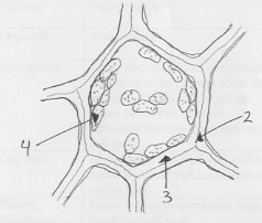
View of the leaf of and Ahacharis plant magnified 400 times
Observations in this drawing:
View of Human cheek cells magnified 400 times:
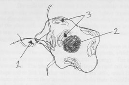
Observations in this drawing:
Plant Cell vs. Animal Cell Comparison Chart
Based on samples of Anacharis cells and Human cheek cells
|
|
Plant Cells |
Animal Cells |
|
Nucleus |
None visible |
Clear, dark nucleus visible and unmoving in the center of the cell |
|
Organells |
None visible |
Unclear, light colored organells were visible. |
|
Cloroplasts |
Many clearly defined cloroplasts were seen to be moving about the cell individually and in groups. There seemed to be some attraction between them and the cell wall. |
None visible |
|
Cell wall |
A very clear cell wall was visible which included various divisions within itself. |
No cell wall |
|
Plasma Membrane |
Possible plasma membrane seen inside the cell wall. |
A very thin plasma membrane was seen surrounding the cell. |
|
Shape |
Boxy, often hexagonal, with nearly strait edges. Did not overlap other cells. |
Bubbly, with no regular shape. Often overlapped other cells. |
Discussion
The cells I observed during this laboratory exercise presented a clear contrast between those cells of the Kingdom Anamalia and those of the Kingdom Plantea. While the cells of both of these kingdoms are classified as eukaryotic and therefore have certain characteristics in common, their functions dictate that they will also be very different.
The cells of both of these kingdoms contain certain organells. In the animal cells these were clearly visible as a nucleus and other lighter organells, and in the plant cells these included the cloroplasts. Both animal and plant cells displayed some sort of cell membrane. In the case of the plant cells this was mostly hidden by the more prominent cell wall, and in the animal cells it was only observed as a very thin line, but in both cases it was there and it did hold the cell together.
However, for all these things these cells have in common, they also have their differences. The plant cells displayed cloroplasts, which contain the plant's chlorophyll allowing it to turn the energy of the sun into usable energy. The animal cells naturally were lacking these cloroplasts. The plant cells also displayed a rigid cell wall which allowed them to maintain their shape and to link together with other plant cells, much like the bricks of a wall. The cell was of plants gives them their strength, but limits their flexibility. This is why the skin of animals is soft and pliable, while the bark of trees is not. Animal cells displayed a clear nucleus which the plant cells did not. This is perhaps misleading, as we do know that plant cells have a nucleus.
In general I believe my observations were compatible with those of the rest of the class. It is possible that a few of the other members of the class were able to see the various aspects of the plant cell wall more clearly or the organells of the animal cell more clearly, but this would not have significantly changed my conclusions.
My prediction that the cells of the Plantea Kingdom would appear boxy, in hexagonal patterns, with thick cell was correct, other than the fact that not all of the cells presented a perfect hexagonal shape. I further predicted that the cells of the Anamalia Kingdom would appear less rigid and more bulbous in shape what lends themselves to the greater variety of movement needed for the members of this kingdom. This prediction was correct.
It was unfortunate that I was unable to view the animal cell with a higher powered microscope that would have allowed me to inspect the various organells in more detail. It is altogether possible that this would have given me valuable insight into their various purposes. It was also unfortunate that I was unable to view the cell walls of the plant cells in more detail as I believe that I could have separated the cell membrane and cell wall with some certainty. I also think that with a more powerful microscope I could have viewed the plant cell's nucleus or other organells. The other clear shortcoming of this particular experiment was that it was performed using only one example from each of the kingdoms. To gain a more clear understanding of the structure of the various eukaryotic cells I feel it would be necessary to view specimens from various plants and animals.