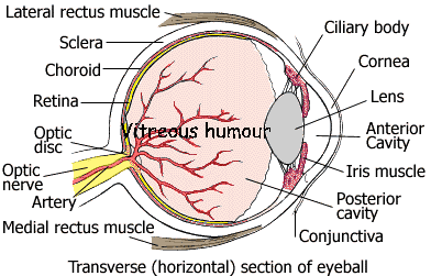
The Structure of EYE

¡@
![]()
| Structures | Functions |
| Anterior Cavity | A cavity of the eye filled with aqueous humor, a fluid that provides oxygen, glucose, and proteins. |
| Choroid | It contains blood vessels which carry nutrients to the eye. It also contains a black pigment which absorbs light rays that enter the eye, preventing their reflection in the eyeball. |
| Ciliary body | It contains the ciliary muscle which controls the thickness of the lens. |
| Conjunctiva | It protects the cornea. |
| Lens | It refracts light rays precisely onto the retina. |
| Cornea | It is transparent. The main function is to refract light rays. |
| Fovea | A depression in the retina where cones (photoreceptor cells) are concentrated and vision is most acute. |
| Iris | The aperture in the centre of the iris is the pupil. Its size is controlled by the circular and radial muscles of the iris. When the circular muscle contracts, the pupil becomes smaller. But when the radial muscle contracts, the pupil becomes larger. |
| Optic disc | Round, flat structure where nerve fibers from the retina converge. |
| Optic nerve | Transmits information about images to the brain. |
| Retina | Contains light-sensitive nerve cells. |
| Sclera | White, outer layer of the eyeball. |
| Vitreous humour | Its functions are to maintain the shape of the eyeball, to refract light rays and to distribute nutrients. |
![]()
¡@