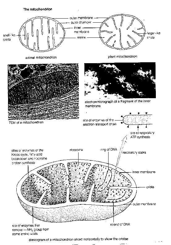
2.The outer membrane is smooth and regular. It controls the entry and exit of
chemicals.
3.The inner membrane is folded inwards to form many tubular processes called
cristae, into the central fluid matrix.These cristae greatly increase the area
for attachment of more respiratory enzymes (electron transport system and
cytochrome system) in a small space.The surface of these cristae has stalked
granules along its length.4.The central cavity is filled with a matrix which is semi-rigid material
containing protein, lipids and trace of DNA. It also contains enzymes for Kreb's
cycle and beta-oxidation of fatty acids
The enzymes (electron transport system )in the mitochondrial cristae and the enzymes of the Kreb's cycle in the matrix are responsible for the oxidative breakdown of food to release energy.Thus mitochondria are present in great number in cells that require a lot of energy, e.g. liver cells, secretory cells.
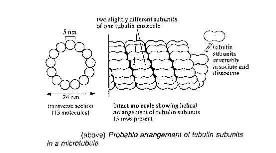
VIII) micro-tubules
Microtubules are elongated, proteinous non-membranous cylinders and are widely distributed in the cytoplasm. They are very fine tubes, with walls made up of helically arranged globular subunits of protein called tubulin. There are three different types of micro-tubules that differ from one another in size, structure and functions:
1. Cytoplasmic micro-tubules they are most abundant near the cell membrane and are usually parallel with cellulose fibres in cell wall. They play a role in cytoplasmic streaming
2. Spindle micro-tubules They are abundant in the nuclear region of the dividing cells and play several roles in cell division.
3. Flagella and cilia Flagella movement results from the sliding of one micro-tubule along another.
XI) Centrioles
Centrioles are two small, dark staining cylindrical bodies which are present in animal cells and certain lower plants. They are adjacent to the nucleus.
(A) Structure
It has 9 peripheral groups of triplet micro-tubules forming a structure of a cylinder. The two centrioles are orientated with their long axis perpendicular to each other.
(B) Function
1. They are concerned with spindle formation during cell division.
2. They are responsible for the formation of cilia flagellum.
X) Nucleus
Nucleus is a spherical body which is bounded by a nuclear membrane and contains nucleolus, chromosomes and nucleoplasm (nuclear sap).
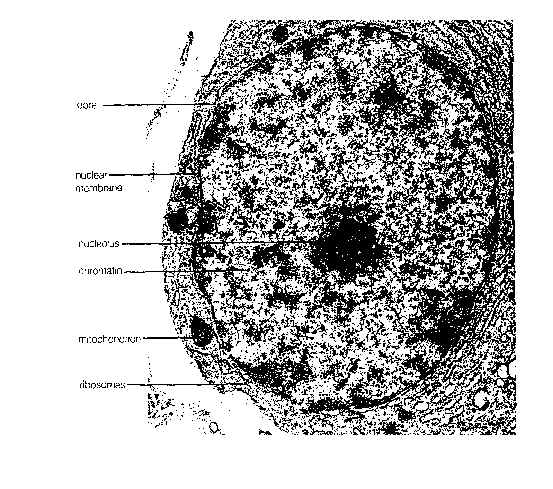
(A) Nuclear membrane(nuclear envelope)
1. It is consists of two membranes. The outer one is continuous with the endoplasmic reticulum and the Golgi bodies so that the nuclear content can be transported to different parts of the cytoplasm and vice versa.
2. It has nuclear pore which control the passage of substances between nucleus and cytoplasm.
(B) Nucleolus
Function: To manufacture rRNA which assembles with protein into ribosomes.It is a dense spherical body. It consists RNA, protein and DNA. The ribosomal RNA is made by the chromosomes and the ribosomal protein may be synthesized in the nucleoli. These two components are assembled in the nucleoli to form ribosomes which subsequently are transported to the cytoplasm where protein synthesis occurs. Most of the nuclear protein synthesis occurs in the nucleoli too. The nucleoli disappeared the early stage of cell division and appear afterwards.
(C) Chromosomes and chromatin
1. Structure
Each chromosome is a rod-like structure which has a constriction, called CENTROMERE, at a definite position along its length. It is the place for the attachment of the spindle fibre and is responsible for its movement during cell division.
When the cell is not in the stage of cell division the chromosome will be in form of many slender thread-like structures. Such diffused state of chromosomes is known as CHROMATIN.However, some remains tightly coiled and continues to stain intensely. This is called HETEROCHROMATIN and is seen as characteristic dark patches near the nuclear envelope.
The remaining, loosely coiled chromatin is located towards the centre of the nucleus and is called EUCHROMATIN. They contain the DNA which is genetically active during interphase.
2. Functions of the nucleus
(a) It contain the genetic material of cell in the form of chromosomes.
(b) The chromosomes contain genes which control the metabolic activities of the cell by controlling the PROTEIN SYNTHESIS that in turn control the synthesis of ENZYMES. Thus the biochemical reactions involved in metabolism can be controlled. ie. nucleus control activities of cell
(c) The chromosomes transmit the genetic information to later generation.
(d) it is involved in the production of ribosomes and RNA.
XI) Cell wall
A) General structure of cell wall
The cell wall has three distinct layers: the middle lamella, the primary wall and the secondary wall.
1. Middle lamella
(a) It is the outermost layer and is shared by adjacent cell.
(b) It consists principally of pectic compounds.
2. Primary wall
(a) It is the middle layer and is the first product of cell wall synthesis by the protoplast.
(b) It contains cellulose microfibrils that are arranged at numerous different angles to one another, giving an interwoven or matted appearance.These microfibrils are separated from one another by some four times their own width. They are embedded in MATRIX made up of pectic compounds(cementing substances) and hemicellulose(A mixed group of alkali-soluble polysaccharides).
(c) It is relatively thin and elastic during cell enlargement. Thickening takes place after completion of cell enlargement and becomes rigid.
3. Secondary wall
(a) It is the innermost layer which is formed by further deposition of pure cellulose on the inner surface of the primary cell wall as the cell matures.
(b) the cellulose microfibrils of the secondary wall are essentially parallel with one another and are bound into macrofibrils. The matrix consists mainly of hemicellulose and in some cells lignin is added.
4. Plasmodesmata(sing.plasmodesma)
(a) The cell wall is penetrated by numerous canals which have an average diameter of less than 0.05 UM.
(b) These canals are filled with fine strand of cytoplasm that connect the adjacent cells together. It also contains cytoplasm and ER.
B) Functions
1. Because of great tensile strength and limited elasticity, they restrain the ballooning and possible rupture of the cells as water diffuses into it.
2. The turgor pressure provides support of the non-woody tissues and organs of plants, whereas the thick walls of cells in wood and sclerenchyma tissues provide mechanical support.
3. The cutinized walls of epidermal cells and the suberized walls of cork cells prevent excessive water loss and desiccation in plants.
XI) Vacuoles
(a) Structure
A vacuole is a fluid-filled sac bounded by a single membrane. Animal cells contain relatively small vacuoles. Plant cells usually have a large central vacuole surrounded by a membrane called the tonoplast. The fluid they contain is called cell sap. It is a concentrated solution of mineral salts, sugars, amino acids, wastes and sometimes pigments.
(B) Functions
1. The sugars and amino acids may act as a temporary food store.
2. The pigments of various colours may colour petals to attract pollinating insects, or fruits to attract animals for dispersal.
3. They as temporary stores for organic wastes. These may accumulate in the vacuoles of leaf cells and are removed when leaves fall.
4.They occasionally contain hydrolytic enzymes and soperfome functions similar to those of lysosomes.
5.They support herbaceous plants, and herbaceous parts of woody plants by providing an osmotic system which creates a pressure potential.
xii) plastids
Plastids, along with vacuoles and cell walls, are structures particularly characteristic of plant cells. Plastids have a double membrane. They are much larger than mitochondria but are less numerous.
Plastids are usually classified on the basis of pigment they contain: leucoplasts(colourless), chromoplasts,(orange or yellow), chloroplast(green)
CHLOROPLAST
(A) Structure
1. Chloroplast is bounded by a double unit membrane,the ENVELOPE, which is smooth and is continuous with no pores or attached particles.
2. The chloroplast always contain chlorophyll and other photosynthetic pigment located on a system of membranes running through a ground substance called STROMA.
3. The membranes system is the site of light reactions in photosynthesis. The membranes are covered chlorophyll and other pigments, enzymes and electron carriers. The system consists of many flattened, fluid-filled sacs called THYLAKOIDS which form stacks called GRANA at intervals, with lamellae(layers) between the grana. Each granum resembles a pile of coins and the lamellae are often sheet-like.
4. The stroma is the site of the dark reactions of photosynthesis. The structure is gel-like, containing soluble enzymes(Enzymes for Calvin cycle), and other chemicals such as sugars and organic acids. Excess carbohydrate from photosynthesis is also stored 'as grains of starch in the stroma. Spherical lipid droplets are often associated with the membranes.
 (B) Function of the lamellae
The lamellae hold the chlorophyll molecules in a suitable position for trapping the maximum amount of light. (Each chloroplast contains 60 grana,, each consisting of about 50 thylakoids. ) The stacking arrangement of thylakoids gives the greatest economy of SPACE. This provides a large SURFACE AREA without taking up too much room.
(C) Function of chloroplast
It is the site of photosynthesis.