interest
Details
of soybean diseases common in the same region as bacterial pustule
(requires Adobe
Acrobat)
Bacterial Pustule of Soybean
Common Name: Bacterial Pustule
Causal Agent: Xanthomonas campestris pathovar glycines, a gram-negative, rod-shaped bacterium
Host Range: This pathovar of X. campestris infects soybeans and some weeds.
Geographic Range: Found wherever soybeans are grown, in warm, wet parts of the world
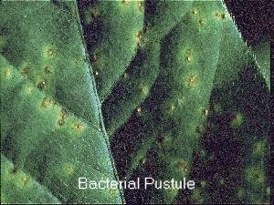
Pustules on a soybean leaf. Note the raised centers
and the
chlorotic halos. (photo courtesy of Michigan
State University)
Symptoms
Soon after infection has taken place, small, light green spots appear on the leaves. These spots become raised in the center; and can appear on both the upper and lower surfaces of the leaf. Raising of these lesions occurs due to rapid growth and multiplication of the cells within the lesion, called hypertrophy and hyperplaisia, respectively. The pustules grow outward and produce necrotic tissue in the center, with a chlorotic halo surrounding the necrotic tissue.
Dissemination
The causal bacteria can overwinter in the soybean seeds themselves or in infected debris. Splashing water and wind can spread the inoculum from the debris onto the soybean plant to facilitate infection. Warm, wet weather aid in the infection process.
Disease Cycle
After the bacterium is placed in contact with the plant, it invades the tissue through natural openings, such as stomata, and through wounds. It multiplies intercellularly and is mobile in the intercellular space until it infects a cell. It then digests part of the cell wall, and binds to the cell. Infected cells experience an increase in cytoplasm, decrease in size and change of shape of chloroplasts, and enlargement of nuclei. These cells collapse and die, and after enough cells have died, tissue dies as well. This dead tissue can harbor the bacteria and facilitate its overwintering.
Control
The best form of control is use of resistant, disease-free seed stock. Crop rotation and burial of inoculum by deep plowing have also been effective in controlling this disease.
References
- Hartman, G.L.; Sinclair, J.B.; and Rupe, J.C. 1999. "Stem Canker." Compendium of Soybean Diseases, Fourth Edition. 6.
- http://cygnus.tamu.edu/Texlab/Fiber/Soybean/sbbp.html Accessed: 4/22/2001.
- Jones, S.B.; and Fett, W.F. 1987. "Bacterial Pustule Disease of Soybean: Microscopy of Pustule Development in a Susceptible Cultivar." Phytopathology. 77:266-274.
In this study, soybeans infected with Xanthomonas campestris were observed using light and electron microscopy to determine how the swelling symptoms develop. Susceptible soybean plants possessing only their first trifoliate leaves were inoculated with the bacteria. The inoculated tissue was sampled at 2 and 7 days after inoculation. Some of the tissue from each sample was then fixed, stained, and observed under a light microscope. The remaining tissue from each of the samples was dehydrated and observed under an electron microscope. In both cases, the slides were photographed under magnification.
The slides showed that pustule development caused by virulent strains begins by stimulating the mesophyll cells near the leaf vein. This takes place when the bacteria eat away slightly at the cell wall and bind to the plant's cell. An increase in cellular cytoplasm and the size of nucleus is experienced, as well as a decrease in size of the chloroplasts and the vacuole. Infected cells eventually burst. Avirulent strains that cause a hypersensitive response are tied up and made inactive by the plant.
Brent Hulke