Advanced Research and Therapeutic Institute ENCEPHALOS
3 Rizariou Street, Halandri 15233, GREECE
Clinical MRI
By E.D. Gotsis,
Ph.D.
A Siemens Magnetom 63/84 SP is the MR imager of our Institute with everything on it, including Proton and Phosphorus-31 MR Spectroscopy, CISS (Construction Interference in the Steady State), 3D Spine Myelography, etc.
In this page we will show a few representative images that depict the quality of our laboratory in mostly non-routine examinations.
An example of CISS (imaging the semicircles of the inner ear) is shown below. Originally two datasets of images are acquired with isotropic resolution of 0.7x0.7x0.7 mm. A standalone program creates a new datase of images based on the original two datasets (4 minutes total acquisition time). Finally the Maximum Intensity Projection (MIP) algorithm is used to reconstruct projections at the desired levels.
CISS imaging of the semicircles
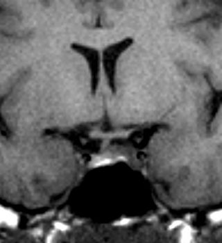
The following two images show why MRI is the modality of choice for imaging the pituitary gland (T1-weighted images before and after Gd-DTPA injection).
 |
 |
| T1-weighted image before GD-DTPA injection | T1-weighted image after Gd-DTPA injection |