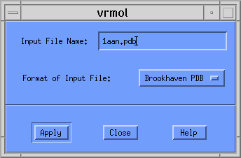
Once this is entered, you should click on the [Apply] button. Another dialog window appears asking you for the type of plot you want. Your choices are
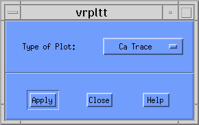
Since PDB structures can be very large, this example generates a Ca Trace plot. This option should be selected from the pull-down menu so that the window looks like the following:
When you click the [Apply] button, VRMol writes the file 1aan.pdb.Ca.wrl to disk. If you had selected Ball and Stick, CPK, or Rods plot, VRMol would have created 1aan.pdb.B-S.wrl, 1aan.pdb.CPK.wrl, or 1aan.pdb.Rods.wrl, respectively.
To see this VRML image from a browser that supports a VRML Plug-in, simply click on 1aan.pdb.Ca.wrl.
Unfortunately, browsers under AIX do not support such a plug-in, and you will have to examine this structure using another product. A few are available, and for ease of use I would recommend an older viewer called VRWeb. For further information, please consult Installing and Using VRWeb.
Program Limitation
The current version of VRMol only plots a single polypeptide chain. This
means that if the PDB structure contains two or more chains, only the first
will be displayed. If you want to display multiple chains, you need to edit
the PDB file and remove the "TER" card between chains. For this
Ca Trace, an erroneous bond will be drawn connecting the last
alpha-carbon of the preceeding chain to the first alhpa-carbon of the
following chain.
� 1999 Brian T. Luke, Ph.D.