Contents and links:
Helga's retina
book:
Ron Douglas:
The primate retina is a bit more complex than the retina of a turtle. In general the cells are much smaller and more densely packed. This makes it substantially more difficult to investigate the morphologic features of these retinas which is one of the reasons why so much research is done on cold blooded species.
Still, many features of the monkey retina bear a striking similarity
to the turtle retina (hope my colleagues will forgive me for making this
statement)
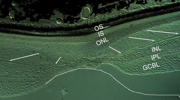 |
| Frozen section of the monkey retina through the fovea region. OS: photoreceptor
outer segments; IS: photoreceptor inner segments. ONL: outer nuclear layer
(cell bodies). INL: inner nuclear layer. IPL: inner plexiform layer. GCBL:
ganglion cell body layer.
In the central retina, the photoreceptors are packed so densely (in several rows on top of each other) that their axon terminals which have to be arranged in a single row (the little spheres at the tips of the arrows) cannot be fit into the same amount of space. As a consequence, the photoreceptors themselves have to have long axons, the course of which is given by the white arrows. Note that a similar arrangement is maintained as discussed earlier for the case of the retinal bipolar cells in the turtle. Also, note that the ganglion cell body layer consists of about 7-8 rows of densely packed retinal ganglion cells. The fovea is the weakest spot within the retina and prone to a variety of diseases as "macular holes" or "macular degeneration", simply because of its thinness. At the same time, this also caused the particular section shown above to be distorted during sectioning. The white line gives the approximate surface outline as it should have been. |
Compared to the foveal region, the peripheral retina looks rather simple. The cells are larger and less complex, there is only one row of photoreceptor with mixed rods and cones, only one layer or retinal ganglion cells which are not even packed very densely.
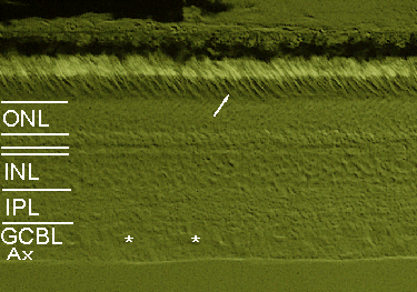 |
| Section of the monkey retina approximately 10 mm away from the fovea (midperiphery of the eye) some of the retinal ganglion cells are marked by an asterisk. the arrow points at a cone, whereas most of the other photoreceptors are rods easily recognized by the light appearance of their outer segments. Note that the first layer indicated below the ONL is the row of photoreceptor axon terminals, i.e. cone pedicles and rod spherules (to name them correctly). Ax: Axons of retinal ganglion cells. |
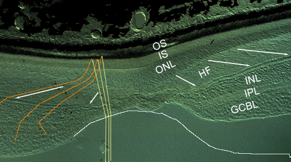
O.k., back to the retinal vs cortical magnification factor in monkey or human. Below, the path of a few light beams is superimposed on the monkey fovea from above (yellow lines). At the vitreo-retinal interface the beams are refracted away from the center of the fovea until they hit the photoreceptors. The orange lines show the further destiny of the (now neuronal) signals. The first step involves a slight bending towards the periphery on the level of photoreceptor inner segments and cell bodies. The major distortion occurs on the level of the photoreceptor axons (Henle's fibers; HF) that transmit the signal to the terminals (cone pedicles) at the point of the arrows. The next level of distortion occurs on the way through the INL, based on the architecture of the bipolar cells. The last step in this regard involves the dense packing of the retinal ganglion cells. since each retinal ganglion cell occupies the same space in the brain, the stacking of these cells into multiple rows alone will already result in a dramatic over-representation but it is the entire retinal architecture which is responsible for the retinal magnification factor in its full extent.
 |